
For patients with scoliosis, the diagnosis is made by physical examination and X-rays. So, when do you need an MRI? In this article, I will try to answer the question of ‘when should an MRI be performed for scoliosis patients’.
Scoliosis mostly occurs without an underlying cause (idiopathic) (see: scoliosis classification). In this idiopathic group, MRI is not routinely performed.
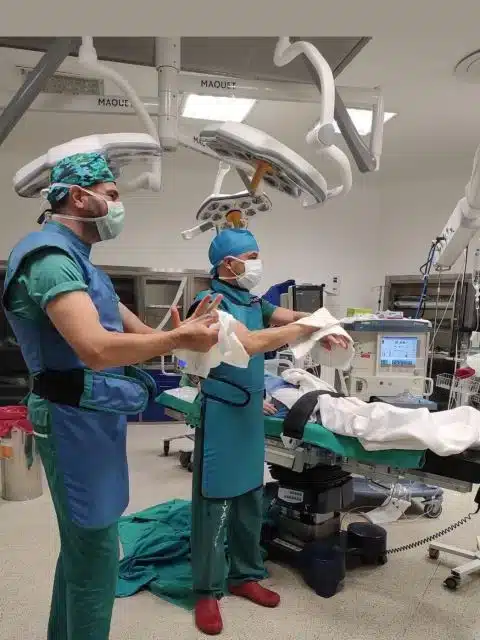
However, in patients who are scheduled for surgery, an MRI should be performed to investigate whether there is any additional pathology in the neural structures before surgery.

Apart from this, the situations that require MRI can be summarized as follows:
Congenital Scoliosis
Congenital scoliosis occurs due to some congenital anomalies of bone and nerve tissue. In order to understand these anomalies completely, they should be investigated with MRI.
Spinal Dysraphism
Spinal dysraphism is the partial fusion or misalignment of the bone and neural structures of the spine due to impaired closure of the neural tube during the development of the embryo. The posterior part of the spine is not completely closed along the midline. Myelomeningocele, neurofibromatosis, achondroplasia, Klippel Feil Syndrome and some connective tissue diseases are in this group.
Patients Under 10 Years of Age
In clinical studies, the probability of finding anomaly in neural structures in scolioses (infantile and juvenile type scoliosis) seen in patients aged 10 years and younger was higher than those over 10 years of age. However, the conclusion that ‘all patients under the age of 10 should undergo MRI’ is not correct. Some criteria, such as the type and size of the curvature, and the presence of findings suggestive of neural anomalies, are effective in deciding for MRI.
Idiopathic Scoliosis with Atypical Features
Some findings in idiopathic scoliosis may be clues for underlying neural anomalies. These findings:
- Male gender
- Pain (especially head and neck pain)
- Abnormal neurological symptoms
- Atypical curves in graph
- Left-sided curvatures
- Short segment curvatures
- Very rapidly progressive curvatures
Clinical Findings
The patient’s complaints and physical examination findings may also be a warning for MRI. These findings, which should make the clinician suspicious of the presence of neural anomalies, are:
- Neurological symptoms (e.g. headache, neck pain)
- Neurological findings (e.g. abnormal superficial abdominal reflex)
- Foot deformities (e.g. Pes cavus)
- Pain
Radiographic Findings
Scoliosis radiographs are those that are taken standing up and allow us to evaluate the entire spine, from the chin to the pelvis. Some abnormal findings on these radiographs may require an MRI:
- Atypical curves (Left-sided or short segment sharp-angle curvatures)
- Sharp thoracic kyphosis
- Rapidly progressive curves
- Presence of dural ectasia (wide canal and thin pedicles)
In summary, these are the situations in which MRI should be performed in scoliosis patients.
Please keep in mind that these criteria are not for our patients to decide on their own about MRI, but to inform you when to have an MRI when you go to the doctor because of scoliosis.