
Congenital scoliosis is scoliosis that occurs due to formation or differentiation defects in the baby’s vertebrae while still in the mother’s womb.
Therefore, the baby has scoliosis at the time of birth.
The developmental defects mentioned here are that vertebra takes a triangular shape instead of a rectangular shape (formation defects) because it cannot be formed completely, or vertebrae cannot be separated from each other and remain conjoined(differentiation defects).
Causes of Congenital Scoliosis
Genetic transmission and some environmental factors are blamed among the causes of congenital scoliosis, but in fact, there is no clear cause.
Although the diagnosis of scoliosis is usually made in the walking age, sometimes it may not be noticed until adolescence.
When describing scoliosis, the deformity of the spine or its relationship with other vertebrae are taken into account. Depending on the type of problem, scoliosis (right or left), kyphosis (hunchback) or lordosis (dimpling) may occur in the child.
However, these curvatures are not usually seen alone, the most common form is kyphoscoliosis, where scoliosis and kyphosis are combined.
There are two reasons for the curvature in congenital scoliosis:
In formation defects, the spine, which should normally be rectangular in shape, becomes triangular because it cannot fully form. As the growth continues, the curvature begins to form or proceeds depending on the structure of this triangle.
In differentiation defects, it is observed that the vertebrae, which should normally be completely separated by disc tissue, remain conjoined on one side. In this case, there will be no growth from the conjoined part but growth will continue from the separate part, and the spine will gradually curve to one side.
These anomalies can be seen in more than one spine and even simultaneously in different types. Since the dorsal vertebrae are attached to the ribs, existing defects can also affect the ribs. As the curvature progresses, the ribs may also begin to bend or rotate, causing the rib cage to deform gradually.
Radiological Examinations
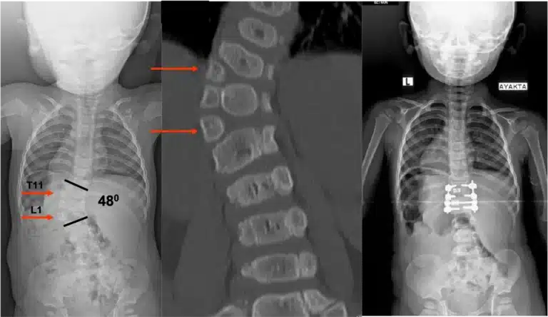
The primary radiological examination for diagnosis is X-ray, the location and shape of the anomaly can be detected in the X-ray.
However, the main assessment is made with computed tomography (CT). The shape and structure of the bone, the size and severity of the defect can be fully revealed by CT.
Besides, Magnetic resonance (MR) should also be taken to evaluate the spinal cord.

Congenital scoliosis can often be accompanied by anomalies of other organs. Since the heart and kidneys are formed at the same time as the spine in the womb, cardiac anomalies (25%) and kidney anomalies (10%) can be seen.
In addition, problems related to the skeletal system such as arm and leg anomalies may accompany.
Therefore, radiological examinations should be performed not only for scoliosis, but also for accompanying anomalies, if any.
Diagnosis and Treatment
The treatment course of congenital scoliosis depends on the age of the child at the time of diagnosis.
The spine grows rapidly in two periods. The first of these is the first five years, and the second is the adolescence period. Therefore, the curvature will progress rapidly during these periods.
In these intervals, curvature should be followed much more closely.
This course of progression will be determined by your doctor, perhaps followed only by observation. Being under observation does not mean that no treatment has been carried out.
After scoliosis and accompanying problems have been identified and if the curvature does not progress, it is possible for the person to continue her/his life without additional treatment.
Corset or plaster cast treatment is not successful for advanced curvatures.
Here, the surgical treatment method is determined by considering the severity of the anomaly, the direction and size of the curvature, the age of the child and the growth potential.
Among the surgical treatment options; there are methods such as freezing the curvature as it is (in situ fusion), freezing(fusion) one side of the spine (hemiepiphysiodesis), removing the half-defective spine (hemivertebra resection), directing growth with growing rods, and VEPTR that can correct the curvature and constriction in the rib cage together.