Our spine consists of bones and discs in between. The task of the spine is to protect the spinal cord and the nerve roots coming out of the spinal cord, to keep the trunk in an upright position and to allow movement.
The discs, located between the bones called vertebrae, consist of two main structures:
In the middle, there is an area with a more liquid consistency called the ‘nucleus’ and is fundamental in carrying the load.
Around the nucleus, there is a second structure called the ‘anulus’, which ensures the integrity of the disc. The annulus is made up of fibers and surrounds the nucleus, locking it where it should be.
The formation of a tear in this annulus structure and the protrusion of the nucleus content is called ‘lumbar hernia’ or ‘herniated disc’.
The resulting hernia causes complaints by pressing on the spinal cord and nerve roots.
There are 6 bones in the lumbar region and 5 discs between them.
Especially the lower part of the waist is the junction point of the body. It bears the weight of the body, but also has to move under a heavy load.
Due to these strains, lumbar hernias are mostly observed in the lower parts of the waist.
Causes and Symptoms of Lumbar Hernia
There are a number of reasons that predispose to lumbar hernia.
These reasons; inactivity, being overweight, making sudden movements that force the waist and being exposed to heavy loads.
The first symptom is usually low back pain.
However, in 80% of low back pain, the underlying cause is not herniated disc. In other words, every low back pain should not be perceived as a hernia.
As the pressure on the nerve increases, numbness and loss of strength also begin to occur.
Much more severe symptoms such as paralysis, urinary and stool incontinence may occur in the future.
Diagnosis of Lumbar Hernia
The diagnosis of herniated disc is made by physical and radiological examinations.
The most important radiological examination is magnetic resonance (MRI).
The location of the hernia, its size, and the severity of the pressure on the nerve are determined.
EMG (nerve measurement) may also be required to see how much the nerve is affected by the compression.
Treatment is guided in the light of these findings.
Treatment Modalities of Lumbar Hernia
Substantially, lumbar hernia can be treated conservatively, that is, without surgery. At this point, the most important deciding factor is whether there is a state of paralysis or not.
If weakness has started in some muscle groups due to the pressure on the nerve, the treatment is surgical.
The aim of surgical treatment is to remove this pressure as soon as possible and to prevent a further paralysis condition.
However, many patients undergo surgery because they cannot stand the pain.
Conservative treatment consists mainly of medication, rest and physiotherapy.
Hernia is often accompanied by contraction of the lumbar muscles. This is because the body reflexively wants to keep itself in a pain-free position. This constant state of contraction adds to the pain caused by the hernia, making the situation worse.
The goal of conservative treatment is to relax and strengthen these muscles and to overcome the pain attack without any problem.
In hernias that do not require surgery but do not respond to conservative treatment, ‘epidural or transforaminal injection’ is performed. The aim here is to reduce this edema and relieve the pain by applying local steroids to the nerve that is crushed under pressure and develops edema.
This procedure can be performed under sterile conditions by imaging the spine with fluoroscopy.
Surgical treatment, on the other hand, is performed through an incision about 2 cm in length.
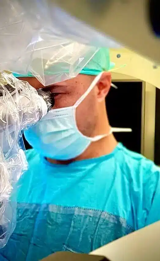
This operation, also known as the closed method, is performed under a microscope. The part that is the cause of the hernia is removed and the nerve under the pressure is relieved.
The patient is discharged within 24 hours and it is recommended to stay away from activities that will force the waist for 3 weeks.
